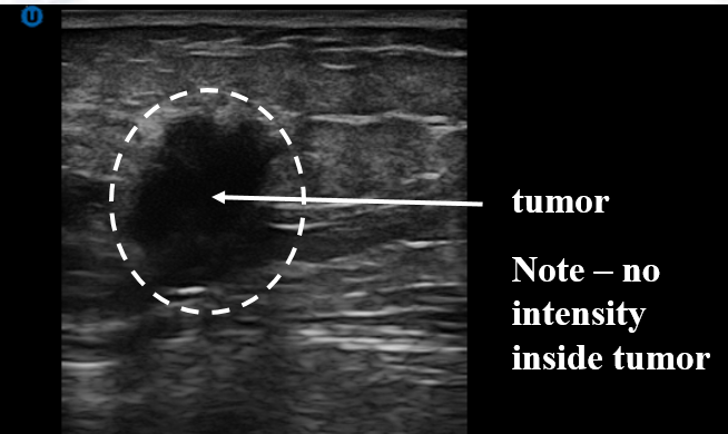
Patient is sitting up for DACI examination. A tumor is identified (arrowed) within the white dashed circle. There is intensity within the tumor area so a diagnosis is possible without an invasive biopsy to determine if it is cancer or benign.
DACI Ultrasound and DACI RAFA Lens Images of Breast Cancer
DACI’s Ultrasound Image
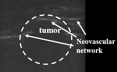
DACI’s RAFA lens Image
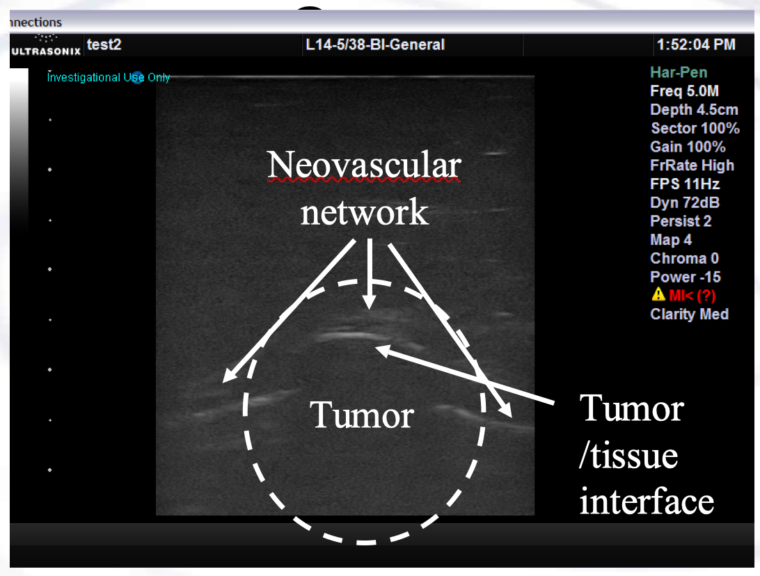
The same breast cancer tumor is imaged using DACI’s Ultrasound and DACI’s RAFA lens showing complimentary information. DACI US sees whole body of tumor whereas DACI RAFA sees neovascular network and measures SOS of tumor.
Speed-of-Sound of Breast Tissues
Previous prototype of DACI successfully measured speed of sound of malignant breast tissue, 1548 m/s*.
* – Fryer, M.J., B.D. Sawicka, P.B. Jacquemin, C.M. Ludgate, P. Howard, W. Beckman, B. Nelson, and R.A. Herring, “Cancer Diagnosis, Treatment and Its Monitoring Using An Acoustic Confocal Holography Microscope,” CSME FORUM 2010, (2010).